• 972-499-3593
    Eminent Spine
    • Timeline
    • The Sword of Eminent
    • Products
          • 3D Titanium Pedicle Screw System
          • Scoliosis Deformity
          • Cervical Implants
            • Cervical Stand-Alone
            • Cervical Plate
            • Cervical IBFD
          • SI Systems
            • Lateral SI
            • Posterior SI
          • Lumbar Systems
            • ALIF Stand-Alone (3D Titanium)
            • ALIF (PEEK)
            • Pedicle Screw
            • 3D PLIF & TLIF
            • PLIF
            • TLIF
            • LLIF
            • Buttress Plate
          • Prep Sets
            • SI Prep Sets
            • Cervical Stand-Alone Prep Sets
            • Cervical Plate Prep Sets
            • ALIF Prep Sets
            • PLIF/TLIF Prep Sets
            • Pedicle Screw Prep Sets (Open & MIS)
          • Kyphoplasty
          • Biologics
    • Resources
      • Surgical Technique Manuals
      • White Papers & Usage Reports
      • IFU’s
      • Blog
    • Case Studies
    • Contact
    • Menu Menu

    A Guide to SI Joint Pain: Why It’s Underdiagnosed and How Surgical Fixation Helps

    Despite advanced imaging technologies, SI joint dysfunction often hides in plain sight, evading CT and MRI scans and leaving patients without answers until a careful physical exam, detailed history, and targeted diagnostic injections finally reveal the true source of their pain.
    Understanding SI Joint Pain

    The sacroiliac (SI) joint, located where the sacrum meets the iliac bones of the pelvis, plays a crucial role in transferring forces between the spine and lower body. Although small, it bears immense biomechanical stress every day. When inflammation, degeneration, or instability affects this joint, the result can be chronic SI joint pain, a condition often mistaken for lumbar spine or hip pathology.

    Studies suggest that SI joint dysfunction accounts for up to 25% of lower back pain cases, yet it frequently goes unrecognized. Because symptoms overlap with other musculoskeletal issues, patients often endure months or years of ineffective treatment before receiving the correct diagnosis.

    Why SI Joint Dysfunction Is Underdiagnosed

    The challenge in diagnosing SI joint pain lies in its nonspecific presentation. Patients typically report pain in the lower back, buttocks, or posterior thigh, regions also affected by lumbar disc herniation or hip osteoarthritis. Even imaging can be inconclusive; radiographs and MRIs may not clearly reveal inflammation or instability at the SI joint.

    Overlapping Symptoms

    • Dull, aching pain radiating to the lower back, hip, or groin
    • Increased discomfort after prolonged standing, sitting, or climbing stairs
    • Asymmetrical pain or tenderness near the posterior superior iliac spine

    Because these symptoms mimic other spinal or hip disorders, SI joint dysfunction often becomes a diagnosis of exclusion. Clinicians may focus on the lumbar spine first, delaying appropriate care.

    Limitations of Imaging Alone

    Standard imaging doesn’t always correlate with pain. Even advanced modalities like CT or MRI may miss subtle joint changes or early instability. Consequently, diagnostic precision depends on a combination of physical assessment, patient history, and confirmatory injections that temporarily relieve pain when the SI joint is anesthetized.

    How SI Joint Pain Is Diagnosed

    Accurate diagnosis typically requires a structured, multi-step approach:

    • Physical Examination: Provocative maneuvers, such as the FABER or Gaenslen test, help reproduce pain associated with SI joint dysfunction.
    • Diagnostic Injections: Fluoroscopically guided injections of local anesthetic confirm the joint as the pain generator when relief exceeds 75%.
    • Elimination of Other Causes: Ruling out lumbar radiculopathy, spinal stenosis, and hip pathology ensures diagnostic accuracy.

    This comprehensive evaluation allows clinicians to identify true SI joint dysfunction and tailor treatment accordingly.

    Conservative Treatments for SI Joint Pain

    Before surgery is considered, most patients begin with non-operative therapies aimed at reducing inflammation and improving joint stability. Common options include:

    • Physical Therapy: Focused on core strengthening and pelvic stabilization exercises.
    • Medications: NSAIDs and corticosteroid injections to control inflammation and pain.
    • Bracing: SI belts or orthoses can temporarily reduce movement and relieve symptoms.

    While conservative care helps many, some patients continue to experience debilitating pain that limits function and quality of life. For those individuals, surgical fixation or fusion may offer lasting relief.

    When Surgery Becomes the Solution

    Surgical treatment for SI joint dysfunction is typically reserved for patients who:

    • Experience persistent pain despite 6+ months of conservative management
    • Have confirmed SI joint pathology through diagnostic injections
    • Show functional limitations affecting daily activity or mobility

    Advancements in SI joint fixation and fusion techniques have made surgery more predictable, less invasive, and more successful than in the past. These procedures aim to stabilize the joint, promote fusion, and eliminate the source of motion-related pain.

    Learn how 3D printed pedicle screw design improves strength, osseointegration, and precision.

    Learn More

    What Is SI Joint Fixation?

    SI joint fixation involves implanting one or more devices across the sacroiliac joint to limit abnormal motion and allow bone fusion. Using minimally invasive surgical approaches, the surgeon inserts specialized implants, typically made from titanium, through a small incision under fluoroscopic guidance.

    The implants achieve stabilization through a combination of:

    • Mechanical fixation: Immediate joint stabilization to reduce micromotion
    • Biologic fusion: Encouraging bone growth across the joint for long-term stability

    This approach reduces recovery time, minimizes tissue disruption, and allows many patients to resume light activity within weeks.

    How SI Fusion Implants Work

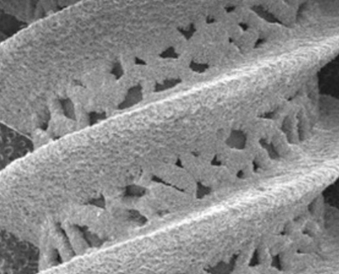
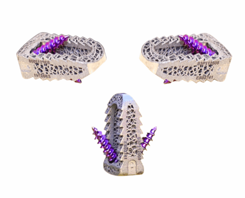
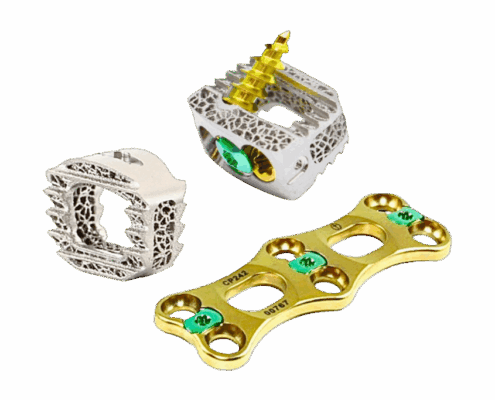
    SI fusion implants are designed to provide both structural integrity and biological integration. Many modern systems, including those from Eminent Spine, utilize porous titanium and 3D-printed architecture to mimic cancellous bone, encouraging rapid osseointegration.

    Key design features include:

    • Surface Porosity: Promotes bone growth and long-term fixation
    • Optimized Thread Geometry: Enhances grip within dense cortical bone
    • Anatomical Sizing: Accommodates a range of patient anatomies and surgical techniques

    Together, these characteristics lead to faster fusion, stronger stability, and more predictable outcomes, both intraoperatively and postoperatively.

    Is SI Joint Pain Surgery Effective?

    Clinical studies consistently show that minimally invasive SI joint fixation provides significant pain relief and functional improvement for appropriately selected patients. Reported benefits include:

    • Reduced pain scores by more than 50% within six months post-op
    • Improved walking tolerance and sitting comfort
    • Decreased dependence on pain medications

    While individual outcomes vary, success largely depends on proper diagnosis, surgical technique, and implant design. For patients with confirmed joint dysfunction, fixation offers one of the most durable solutions available.

    The Future of SI Joint Surgery

    The increasing recognition of SI joint dysfunction is reshaping how spinal and orthopedic surgeons approach chronic low back pain. As awareness grows, additive manufacturing and implant innovation will continue to refine surgical options, offering even more effective solutions for long-term stabilization and pain relief.

    With continued advancements in material science, imaging, and surgical navigation, SI joint fixation will likely become a standard component of comprehensive spinal care—providing patients with durable, life-changing results.

    Eminent Spine’s SI Joint Solutions

    At the forefront of spinal innovation, Eminent Spine has applied its expertise in titanium additive manufacturing to advance SI joint fusion technology. Our SI implant systems integrate the same engineering principles that define its industry-leading pedicle screw and interbody platforms—strength, precision, and biological compatibility.

    Key Features of Eminent Spine’s SI Fusion System:

    • 3D-Printed Titanium Design: Replicates bone structure for optimal osseointegration
    • Threaded Fixation Profile: Ensures immediate mechanical stability
    • Minimally Invasive Delivery: Reduces operative time and postoperative discomfort

    By combining surgeon feedback with advanced biomaterial engineering, Eminent Spine delivers SI joint fixation systems that support superior outcomes and long-term fusion success.

    Share This Post

    • Share on Facebook
    • Share on X
    • Share on WhatsApp
    • Share on Pinterest
    • Share on LinkedIn
    • Share on Tumblr
    • Share on Vk
    • Share on Reddit
    • Share by Mail

    More Like This

    Revised Screw Posterior Image

    The End of One-Size-Fits-All: How Precision Spine Care Is Reshaping Surgical Outcomes

    https://www.eminentspine.com/wp-content/uploads/2025/11/revised-screw-posterior-image.png 799 1545 Abstrakt Marketing /wp-content/uploads/2025/08/ES-Logo-Edit-2.png Abstrakt Marketing2026-04-07 16:56:302026-05-18 17:19:11The End of One-Size-Fits-All: How Precision Spine Care Is Reshaping Surgical Outcomes
    Cervical Implants Parent Header Image 845x684

    How to Choose a Spinal Implant: A Surgeon’s Guide

    https://www.eminentspine.com/wp-content/uploads/2025/11/Cervical-Implants-Parent-Header-Image-845x684-1.png 684 845 Abstrakt Marketing /wp-content/uploads/2025/08/ES-Logo-Edit-2.png Abstrakt Marketing2026-01-30 03:43:572026-06-23 15:29:20How to Choose a Spinal Implant: A Surgeon’s Guide
    3d Printed Titanium Image

    Comparing 3D Printed Spinal Implants vs Traditional

    https://www.eminentspine.com/wp-content/uploads/2025/08/3D-Printed-Titanium-Image.jpg 500 500 Abstrakt Marketing /wp-content/uploads/2025/08/ES-Logo-Edit-2.png Abstrakt Marketing2026-01-30 03:36:592026-05-18 17:19:11Comparing 3D Printed Spinal Implants vs Traditional

    Categories

    • 3d Pedicle Screw Education & Persuasion
    • 3D Titanium Pedicle Screw
    • Lumbar Fusion
    • Miscellaneous
    • SI Joint Solutions
    • Spinal Implant Education
    • Spinal Implant Innovation
    Es Logo Edit

    Products

    3D Titanium Pedicle Screw System

    SI

    Scoliosis Deformity

    Cervical Implants

    Lumbar Systems

    Prep Sets

    Biologics

    Resources

    Surgical Technique Manuals

    Case Studies

    White Papers & Usage Reports

    IFU’s

    1-2 Page Product Information

    Contact

    2004 Ventura Dr. Ste 250
    Plano TX 75093

    972.499.3593

    [email protected]

    Website by Abstrakt Marketing Group ©
      • Sitemap
      • Privacy Policy
      Link to: Comparing 3D Printed Spinal Implants vs Traditional Link to: Comparing 3D Printed Spinal Implants vs Traditional 3d Printed Titanium ImageComparing 3D Printed Spinal Implants vs Traditional
      Scroll to top Scroll to top Scroll to top

      This site uses cookies. By continuing to browse the site, you are agreeing to our use of cookies.

      AcceptLearn more

      Cookie and Privacy Settings


      How we use cookies

      We may request cookies to be set on your device. We use cookies to let us know when you visit our websites, how you interact with us, to enrich your user experience, and to customize your relationship with our website.

      Click on the different category headings to find out more. You can also change some of your preferences. Note that blocking some types of cookies may impact your experience on our websites and the services we are able to offer.

      Essential Website Cookies

      These cookies are strictly necessary to provide you with services available through our website and to use some of its features.

      Because these cookies are strictly necessary to deliver the website, refusing them will have impact how our site functions. You always can block or delete cookies by changing your browser settings and force blocking all cookies on this website. But this will always prompt you to accept/refuse cookies when revisiting our site.

      We fully respect if you want to refuse cookies but to avoid asking you again and again kindly allow us to store a cookie for that. You are free to opt out any time or opt in for other cookies to get a better experience. If you refuse cookies we will remove all set cookies in our domain.

      We provide you with a list of stored cookies on your computer in our domain so you can check what we stored. Due to security reasons we are not able to show or modify cookies from other domains. You can check these in your browser security settings.

      Other external services

      We also use different external services like Google Webfonts, Google Maps, and external Video providers. Since these providers may collect personal data like your IP address we allow you to block them here. Please be aware that this might heavily reduce the functionality and appearance of our site. Changes will take effect once you reload the page.

      Google Webfont Settings:

      Google Map Settings:

      Google reCaptcha Settings:

      Vimeo and Youtube video embeds:

      Accept settingsHide notification only